Imaging

See more. Understand more.

Explore imaging with clear information and a thoughtful approach to your visit. Know what to expect before your next step.

Explore imaging services

Illustrative clinical photography · not a VERADX facility

Imaging, with reassurance
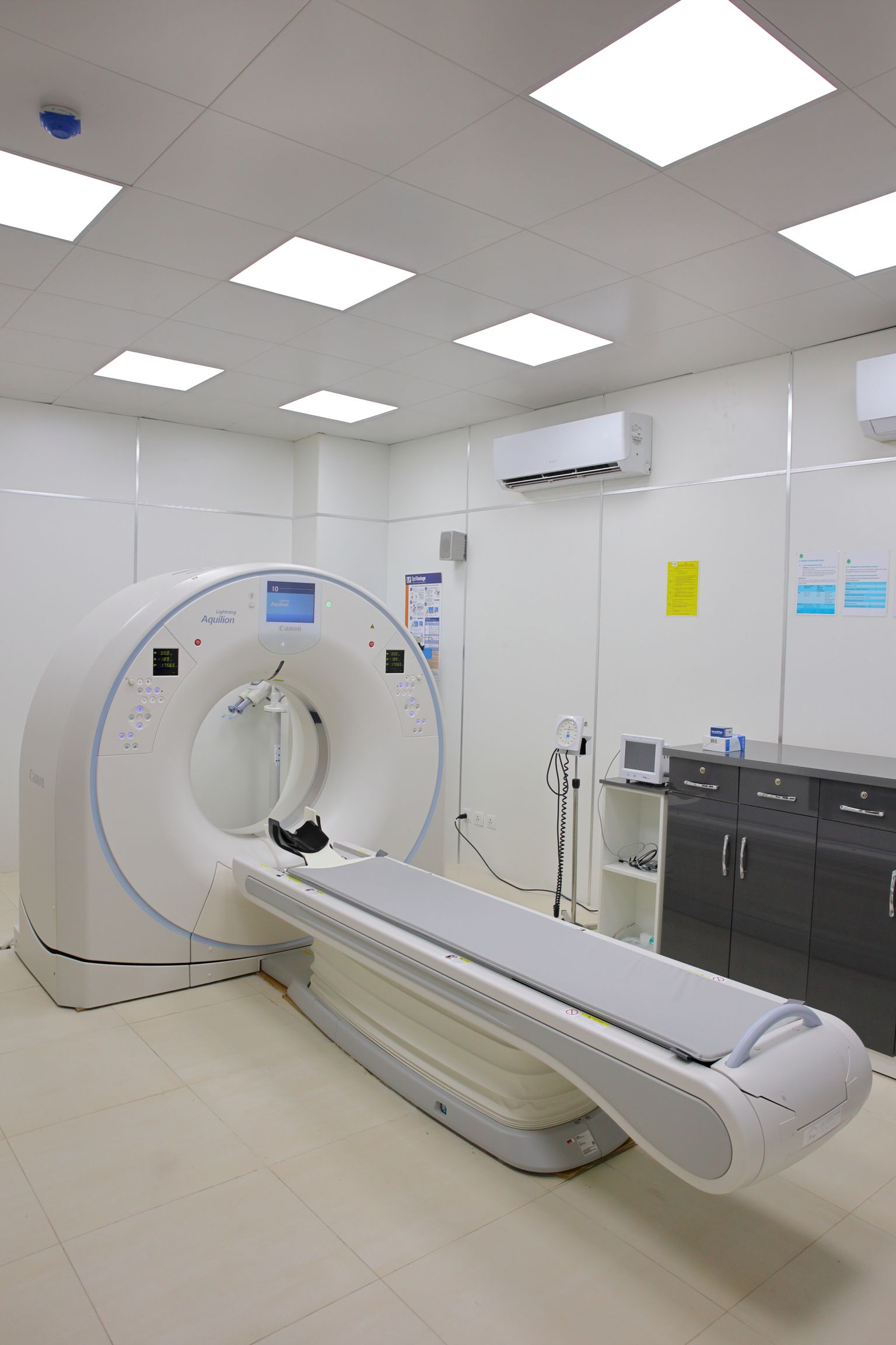
CT scanner with its patient table and controls in a bright clinical room

A clearer picture. A calmer experience.

Science & care / VERADX DIAGNOSTICS

Services

The right information, from the start.

Imaging choice depends on the clinical question and individual circumstances. Times and locations below are illustrative demo information, not live availability. Preparation varies by examination; follow your imaging team’s instructions.

MRI scanner and patient table inside a clinical imaging room
01

Magnetic resonance imaging

MRI

Magnetic fields and radio waves produce detailed images of internal structures without ionising radiation.

Common uses

Brain and spine assessment · Joint and soft-tissue assessment

Preparation

Complete implant and metal screening. Remove metallic items as instructed; eating and drinking instructions depend on the examination.

Tell the team about implants, metal fragments, pregnancy, kidney disease and previous contrast reactions. MRI requires individual safety screening.

Approx. 30–60 minutes
Report: 2–3 working days · demo estimate
London · Central; Manchester · Deansgate
Booking preview availableExplore service & appointment
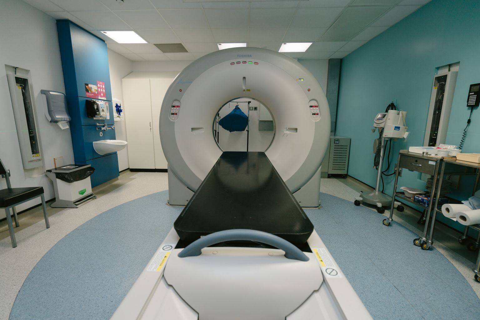
CT scanner in a bright diagnostic examination room
02

Computed tomography

CT Scan

A series of X-ray measurements creates cross-sectional images of the body.

Common uses

Chest and abdominal assessment · Assessment of injuries

Preparation

Ask whether contrast and fasting are needed for your specific scan. Share medicines, allergies and relevant medical history.

Uses ionising radiation. Tell the team if pregnancy is possible and about kidney problems or previous contrast reactions.

Approx. 15–30 minutes
Report: 2–3 working days · demo estimate
London · Central
Booking preview availableExplore service & appointment
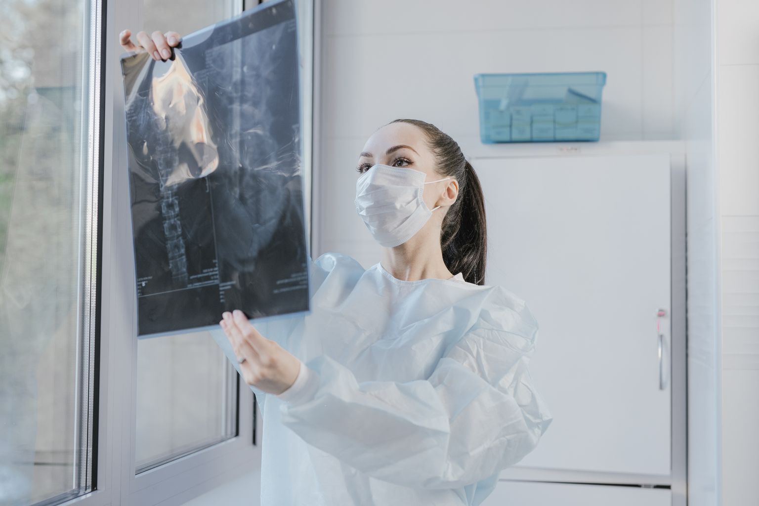
Healthcare professional examining an X-ray film near a window
03

Conventional radiography

X-Ray

X-rays create images that can help clinicians assess bones and other structures.

Common uses

Bone injury assessment · Joint alignment assessment

Preparation

Wear clothing that is easy to change and remove jewellery near the area being examined when asked.

Uses ionising radiation. Tell the imaging team if you are or could be pregnant before the examination.

Approx. 10–20 minutes
Report: 1–2 working days · demo estimate
London · Central; Bristol · Clifton
Booking preview availableExplore service & appointment
Doctor carrying out an ultrasound examination
04

Sound-wave imaging

Ultrasound

High-frequency sound waves create real-time images of structures inside the body.

Common uses

Abdominal organ assessment · Soft-tissue assessment

Preparation

Some examinations need fasting or a full bladder. Follow the instructions for the specific area being scanned.

Does not use ionising radiation. Examination type, including whether an internal scan is proposed, should be discussed with the team.

Approx. 20–45 minutes
Report: 1–3 working days · demo estimate
London · Central; Manchester · Deansgate; Bristol · Clifton
Booking preview availableExplore service & appointment
Mammography equipment photographed by the National Cancer Institute
05

Breast imaging

Mammography

Specialised X-ray imaging examines breast tissue for screening or diagnostic purposes when appropriate.

Common uses

Clinician-directed breast assessment · Screening within an appropriate programme

Preparation

Avoid deodorant, powder or lotion on the breasts and underarms on the day. Bring previous breast imaging if requested.

Uses ionising radiation and breast compression. Discuss possible pregnancy, implants and any symptoms with the team; screening suitability is individual.

Approx. 20–30 minutes
Report: 3–5 working days · demo estimate
London · Central
Booking preview availableExplore service & appointment
Patient undergoing a DXA bone-density examination at the ALSPAC clinic
06

DXA / DEXA scan

Bone Density

Dual-energy X-ray absorptiometry measures bone mineral density, often at the hip and spine.

Common uses

Bone density measurement · Monitoring changes in bone density

Preparation

Tell the team about recent contrast examinations and ask for instructions about calcium supplements before your visit.

Uses ionising radiation. Tell the team if pregnancy is possible and discuss whether the examination is appropriate for you.

Approx. 10–30 minutes
Report: 2–3 working days · demo estimate
London · Central; Bristol · Clifton
Booking preview availableExplore service & appointment
Doctor performing an ultrasound examination with a clinical scanner
07

Vascular ultrasound

Doppler

An ultrasound technique evaluates movement of blood through blood vessels.

Common uses

Blood-flow assessment · Assessment of selected arteries and veins

Preparation

Preparation depends on the blood vessels examined. Ask whether any fasting or clothing instructions apply.

Does not use ionising radiation. The scan and its findings require clinical interpretation; it is not a substitute for urgent assessment.

Approx. 30–60 minutes
Report: 1–3 working days · demo estimate
London · Central; Manchester · Deansgate
Booking preview availableExplore service & appointment
Combined PET and CT scanner with a patient table in a clinical imaging room
08

Functional and anatomical imaging

PET/CT

PET uses a radiotracer to show activity in tissues, combined with CT images of anatomy.

Common uses

Selected cancer assessment and monitoring · Specialist-directed metabolic imaging

Preparation

Follow tracer-specific food, drink and activity instructions. Discuss diabetes and medicines with the team before your visit.

Involves a radiotracer and ionising radiation. Discuss pregnancy, breastfeeding and any contact precautions with the specialist team.

Approx. 2–3 hours including preparation
Report: 3–5 working days · demo estimate
London · Central
Booking preview availableExplore service & appointment

Clinical overview and safety information informed by RadiologyInfo.org. Each detail page links to the relevant patient guide. No imaging is performed or interpreted by this website.

The experience, considered

More than an appointment.
Care at every step.

Speak to our team
Bright clinical corridor with clear routes between examination rooms

01 / Arrival

A calmer beginning.

Plan your arrival and review the details for your chosen centre.

Patient discussing the visit with a doctor in a well-lit consultation room

02 / Your questions

Time to understand.

Discuss preparation, comfort and what to expect with your care team.

Doctor explaining a radiograph to a patient during a consultation

03 / The next step

A clearer conversation.

Learn about the process behind your examination and the information that follows.

Explore the wider catalogue

Different needs. Thoughtful options.

Clinical photography credits

Images are cropped to fit and lightly colour graded. Adapted presentations of the Creative Commons photographs retain their respective licences. Photography illustrates the examination; it does not depict VERADX facilities or current equipment availability.